Most dry eye patients go through a progression of treatments: artificial tears first, then prescription drops, then in-office procedures. For the majority of patients, this approach works — they find a combination that brings their symptoms under control. But for some, the disease has progressed to a point where the ocular surface itself is damaged, inflamed, and struggling to heal. Standard treatments provide only partial relief, and the cycle of discomfort continues.
For these patients, amniotic membrane therapy is one of the most powerful tools available. At Toluca Lake Optometric Center in Burbank, CA, we use amniotic tissue as part of our advanced dry eye care — and the results in appropriately selected patients can be transformative.
What Is Amniotic Membrane?
The amniotic membrane is the innermost layer of the placental tissue that surrounds a developing fetus. It is rich in growth factors, anti-inflammatory proteins, and extracellular matrix components that are essential for tissue healing and regeneration. Because of these properties, amniotic tissue has been used in medicine for over a century — initially in burn and wound care, and more recently in ophthalmic surgery and ocular surface disease.
The amniotic tissue used in clinical treatment is sourced from consented donors, processed, sterilized, and either preserved (non-viable) or cryopreserved (retaining live cells, for maximum biological activity). It contains no viable fetal cells and carries no significant risk of immune rejection.
How Amniotic Membrane Helps the Ocular Surface
When applied to the surface of the eye, amniotic membrane works through several mechanisms simultaneously:
Anti-inflammatory action. The tissue is rich in molecules that suppress inflammatory pathways on the ocular surface. Chronic dry eye creates persistent inflammation that damages the corneal epithelium and the conjunctiva. Amniotic membrane helps interrupt this cycle at a cellular level.
Growth factor delivery. The tissue releases a range of growth factors — including EGF, TGF-beta, and PDGF — that signal damaged surface cells to repair and regenerate. For corneas with persistent epithelial defects, punctate keratitis, or surface erosions from chronic dry eye, this growth factor activity promotes healing that artificial tears cannot.
Physical protection. The membrane acts as a biological bandage over the ocular surface, reducing exposure to environmental irritants and protecting fragile, inflamed tissue while it heals.
Stem cell support. Amniotic tissue supports limbal stem cells — the cells at the edge of the cornea that are responsible for maintaining and renewing the corneal epithelium. In advanced ocular surface disease, limbal stem cell populations can be depleted, impairing the cornea's ability to repair itself. Amniotic membrane supports their function and survival.
When Is Amniotic Membrane Used for Dry Eye?
Amniotic tissue is not the first-line dry eye treatment — it is a specialized intervention for patients whose disease has exceeded what standard therapies can manage. We consider it for patients with:
- Severe, refractory dry eye that has not responded adequately to IPL, prescription drops, punctal plugs, or other treatments
- Corneal staining, punctate epithelial erosions, or persistent epithelial defects
- Ocular surface disease associated with Sjögren's syndrome, graft-versus-host disease, or Stevens-Johnson syndrome
- Significant inflammation that is not controlled with topical treatments alone
- Post-surgical ocular surface compromise
In these cases, treating the surface with biological tissue rather than pharmacological drops can provide a level of repair and relief that no other approach matches.
How the Treatment Is Applied
Amniotic membrane can be delivered in two formats:
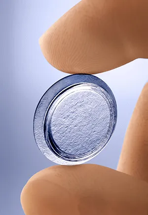
PROKERA is a biologic corneal bandage device that holds a cryopreserved amniotic membrane in a ring. It is placed on the eye like a large contact lens and worn continuously for several days to a week. During that time, the tissue releases its anti-inflammatory and growth-promoting factors directly onto the ocular surface.
Amniotic membrane grafts can also be sutured or glued into position in a clinical setting for patients with more complex surface disease.
The PROKERA format is typically used for advanced dry eye and surface disease and is managed in our office. It is well tolerated by most patients, though vision is blurred while the device is in place.
What to Expect and Who It Helps Most
Patients who undergo amniotic membrane therapy for severe dry eye often describe a meaningful reduction in pain, burning, and light sensitivity during and after treatment. Many experience improved comfort that persists for months following the intervention — not because the underlying disease is cured, but because the surface has been given the opportunity to heal and reset, making ongoing maintenance treatments more effective.
Amniotic membrane therapy is not a replacement for the management of whatever is driving the dry eye — the inflammation, the meibomian gland dysfunction, or the systemic condition — but it can provide critical relief while that management continues.
If you have severe dry eye that has not responded to conventional treatments, contact Toluca Lake Optometric Center in Burbank at (818) 841-1212 to discuss whether amniotic membrane therapy is an appropriate next step for you.