Most people who come to Toluca Lake Optometric Center with chronic dry eye have already been told they have dry eye. They have tried artificial tears. Some have been given prescription drops. A few have seen multiple doctors. What they have not had, in many cases, is a thorough diagnostic evaluation that identifies specifically what is causing their dry eye and what the current state of their tear-producing structures actually is.
The distinction matters enormously. Dry eye is not a single condition — it is a category of conditions with multiple underlying mechanisms. Treating a patient with primarily aqueous-deficient dry eye the same way you treat a patient with predominantly evaporative dry eye driven by meibomian gland dysfunction will produce predictably incomplete results. The treatment has to match the diagnosis.
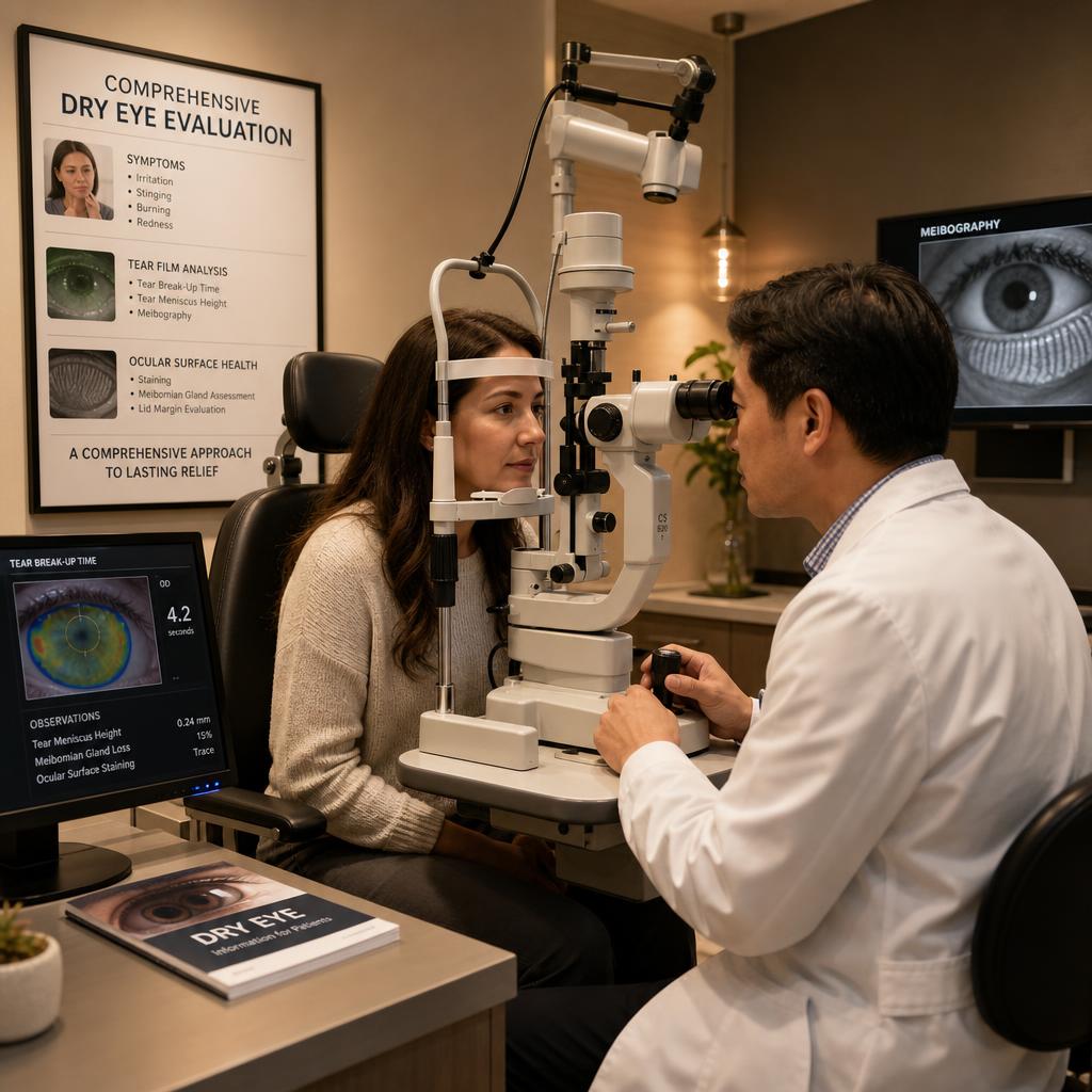
Here is what a comprehensive dry eye evaluation at our Burbank practice includes and what each measurement tells us.
Symptom Questionnaires
The evaluation begins with validated symptom questionnaires — the most common being the OSDI (Ocular Surface Disease Index) or the DEQ-5. These are not just formalities. They quantify the severity and impact of your symptoms in a standardized way that allows us to track your progress objectively over time, compare against population norms, and identify symptom patterns that point toward particular types of dry eye.
Symptom severity does not always correlate with objective findings — some patients with significant gland loss have mild symptoms, while others with less structural damage are very symptomatic. Both patterns matter and influence how we approach treatment.
Tear Film Stability Assessment
Tear breakup time (TBUT) measures how quickly the tear film begins to break apart after a blink. In a healthy eye, the tear film stays intact for at least 10 seconds. In evaporative dry eye, it may break in two to three seconds — or less.
We measure TBUT using fluorescein dye under a slit lamp, or with non-invasive technology that captures tear film images over time. Short breakup time is one of the most direct indicators of meibomian gland dysfunction and unstable tear film lipid layer.
Meibography
Meibography uses infrared imaging to photograph the meibomian glands inside the eyelids. The glands appear as elongated, finger-like structures. Healthy glands are full, clearly defined, and intact. Diseased glands may appear shortened, dilated, irregular, or partially absent — replaced by fat or fibrous tissue as they atrophy.
This is one of the most important tests we perform, because it shows us not just that the glands are dysfunctional, but how much structural gland tissue remains. Patients with significant gland loss require a different treatment approach — and more urgency — than those whose glands are structurally preserved but functionally impaired.
Meibography also gives us a baseline to track. After IPL or other treatments, follow-up imaging can show whether gland structure has improved or stabilized.
Meibomian Gland Secretion Quality Assessment
Beyond structure, we assess what the glands are actually producing. By applying gentle pressure to the eyelid margins, we evaluate whether the glands express any secretion at all, and whether that secretion is clear and oily (healthy) or cloudy, thickened, or waxy (abnormal).
This assessment, combined with meibography, gives us a complete picture of gland health: structure plus function.
Tear Volume Assessment
Schirmer's test and phenol red thread testing measure the aqueous (water) component of tear production. A small paper strip is placed under the lower eyelid and the amount of tear wetting is measured after a set time.
This helps us distinguish between aqueous-deficient dry eye — where the lacrimal gland is not producing enough water-based tears — and evaporative dry eye, where tear volume may be adequate but the tears evaporate too quickly due to inadequate oil. The treatment approach differs significantly between the two.
Ocular Surface Staining
Fluorescein and lissamine green dyes stain damaged cells on the cornea and conjunctiva. Areas of staining indicate active tissue damage from dryness, inflammation, or friction. The pattern and location of staining can point to specific diagnoses — inferior corneal staining from MGD, exposure keratopathy, or superior staining from contact lens-related issues.
Staining severity correlates with the inflammatory burden on the surface and helps guide the urgency of treatment.
Lid and Lash Evaluation
We examine the eyelid margins carefully at the slit lamp — looking for collarettes (Demodex blepharitis), meibomian gland plugging, lid margin thickening, notching, telangiectasias (rosacea), bacterial blepharitis, and any lid position abnormalities that affect tear drainage or distribution.
This examination identifies the underlying lid disease that is often the primary driver of dry eye — the problem that artificial tears can never fix.
Building Your Treatment Plan
With this information in hand, we can tell you not just that you have dry eye, but what type you have, how severe the structural changes are, what is driving it, and what treatments are most likely to actually work for your specific presentation.
That is the difference between managing dry eye and treating it.
Call Toluca Lake Optometric Center in Burbank at (818) 841-1212 to schedule a comprehensive dry eye evaluation.